Futura News

Home > News
que-es-la-miomatosis-uterina-y-como-afecta-tu-fertilidad
Fertility

What is uterine myomatosis and how does it affect fertility?

Uterine myomatosis is the appearance of myomas or fibroids inside the uterus. These are benign tumors classified by their location.  The myomas or fibroids inside a woman’s uterus are classified in the following way, according to their location:   Intramural: located in the uterus muscle wall and can affect fertility if they reach more than 4 centimeters.  Submucosal: formed in the surface of the uterine lining and can affect embryo implantation. Subserosal: located in the outer cover of the uterus and usually doesn’t affect fertility. Is it a common condition? It is the most common pelvic tumor. Approximately 20% of women suffer of uterine myomatosis during their reproductive age (from their first menstruation until menopause) and particularly women ovdf the age of 30.  Uterine myomas can be microscopic or slowly grow until filling the entire uterus. They have different shapes and sizes, and although they could be just one, most of the time are multiple.  How does uterine myomatosis affect fertility? These tumors are present in both fertile and infertile women. Nevertheless, 80% of the cases of infertility because of the uterine factor, is because of the appearance of uterine fibroids that affect the uterine cavity, like the case of intramural and submucosal.  The location of the uterine fibroids is very important because this determines whether they can potentially cause infertility, change sperm flow, or negatively affect the embryo implantation process.  Could this condition affect pregnancy?  Uterine myomas can cause compilations in the pregnancy process, such as:   High risk of miscarriage  Fetal growth retardation Premature delivery because of less space in the uterus Obstruction of the cervical canal  Obstruction of labor  Postpartum hemorrhage What are the symptoms?  Around 25% of uterine myomas are asymptomatic. But they can also manifest through:  Feeling of pressure in lower abs  Abnormally abundant menstrual bleeding  Longer than usual menstrual cycles Painful pelvic cramps Bleeding not related to menstruation  Pain or difficulty while peeing  Infertility Recurrent miscarriages It is important to point out that uterine myomatosis symptoms depend on the size, location and number of present fibroids.  What are the causes?  Although science hasn’t found the specific cause of the uterine myomas, it is known that appearance is related with genetic predisposition.  Also, the growth is associated with hormonal changes in the ovaries, in particular with progesterone and estrogen production.  Uterine fibroids appear during the reproductive stage of women, growing during pregnancy (when there is a high level of hormones), and disappear after menopause when the hormone levels are lower.  How can I get a diagnosis?  Reaching a specialist is the first step. The next one, probably, would be a vaginal ultrasound. Which is the most reliable study for uterine myomatosis diagnosis.  It offers nearly 95% of accuracy when done abdominally and 100% when done intravaginally.  The vaginal ultrasound must be interpreted by an expert. Can I become a mother despite being diagnosed with Uterine Myomatosis?  When it comes to achieving a healthy pregnancy,  treatment of uterine myomatosis depends on the age of the woman, and, above all, the size and location of the fibroids.  Subserosal myomas rarely affect fertility, but patients with submucosal or intramural fibroids larger than 2 inches, are recommended to consider treatments such as In Vitro Fertilization (IVF), especially if they are over the age of 35.  In Vitro Fertilization (IVF) is highly effective when uterine myomatosis does not affect the endometrial cavity.  Fibroids usually change sperm traffic and the embryo implantation process, but In Vitro Fertilization makes it possible for egg fertilization to take place in the laboratory.  Later, the embryo with the highest implantation potential is transferred into the uterus. How do I prepare for the process?  Very rarely do women have to go through a myomectomy before proceeding with the In Vitro Fertilization (IVF) treatment.  This is a surgery to take out the uterine fibroids, after which the pregnancy probability increases and there is an important decrease in the risk of miscarriage.  The new IVF cycle must be performed immediately after the uterine myomectomy because the probability of pregnancy decreases after the first year due to uterine fibroids’ recurrence.  If you would like to learn more about uterine myomatosis, visit our guide: What is Uterine Myomatosis?

Read More »
Assisted Reproduction

Implantation failure

Implantation failure is diagnosed when a baby has not been achieved after at least three cycles of In Vitro fertilization (IVF) or the transfer of more than ten good quality embryos. Currently, there are highly effective solutions for each of the causes of implantation failure, among which the transfer of embryos in the blastocyst phase (day 5 of embryonic development), preimplantation genetic diagnosis and assisted hatching (or assisted hatching). What is the implementation failure? Embryo implantation is a complex process that requires a healthy embryo and a receptive endometrium, as well as a close dialogue between the two. In the field of assisted reproduction, implantation failure is diagnosed when a baby has not been achieved after at least three cycles of In Vitro fertilization (IVF) or the transfer of a total of ten good quality embryos. Causes of implantation failure The causes of implantation failure are multiple and both embryonic problems and problems with the uterine environment may be involved: How are the causes of implantation failure diagnosed? There are several tests that allow us to determine the causes of implantation failure: It is important to note that a detailed evaluation of the cycles carried out so far (number of eggs obtained, fertilization rate, techniques used and embryonic development) is essential to establish an accurate prognosis. Treatments to have a baby after implantation failure Knowing the causes of implantation failure is essential when it comes to selecting a treatment that allows you to achieve a baby with the least wear and tear. Currently, there are highly effective solutions for each of these causes. When implantation failure is associated with immunological problems, these can be easily treated with medications, while if it is due to problems with the uterine environment, treatment varies depending on the cause and can range from hormonal therapy to surgery in very specific cases. However, when implantation failure is due to embryonic problems, the main treatments include: Until a few years ago, all embryos were transferred on the third day after fertilization because it was impossible to keep them alive in a laboratory, but today technology allows us to culture them for a longer time, which makes it easier to select the oldest embryos. quality and with greater power of implementation. (If you want to know more about prolonged embryo culture click here) This technique allows only embryos free of chromosomal abnormalities (aneuploidies) to be transferred to the mother’s uterus. This is related to implantation failure, recurrent pregnancy loss and the woman’s age, since as the woman progresses, the risk of presenting chromosomal abnormalities in the baby increases. This is why transferring only embryos free of these genetic alterations allows us to maximize the chances of implantation, pregnancy and having a baby at home regardless of the woman’s age. Before implantation in the uterus, the embryo must be freed from the membrane that surrounds it (zona pellucida) to come into direct contact with the cells of the endometrium. In some cases, implantation failure is due to the inability of the embryo to leave the zona pellucida and it is necessary to facilitate this process to increase the probability of implantation. At Ingenes we have embryologists who are experts in micromanipulation techniques, which allows us to perform the laser-assisted hatching procedure quickly and accurately without causing any damage to the embryos.

Read More »
Fertility

Uterine Myomatosis How does it affect fertility?

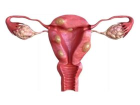
Uterine myomatosis is the appearance of fibroids or fibroids, the most common solid tumors of the uterus in women of reproductive age. They are generally benign estrogen-dependent tumors. They are also called myoma, leiomyoma, or fibroma.Epidemiologically, 2 out of 5 women who present with fibroids do not have any symptoms. They are not a common cause of infertility. For patients who have fertility problems, fibroids have a prevalence of 5-10%. Only in 1 to 2.5% of cases is it a cause of infertility. (1) Among the risk factors that contribute to the development of uterine fibroids are nulliparity, black race, obesity, genetic factors, early menarche, alcohol, and caffeine. (2) The classification of uterine fibroids is based on the relationship they have with the uterine wall, thus they can be subserosal, intramural, and submucosal. 95% of fibroids are located in the body of the uterus, and only 5% are located at the neck. Subserosal fibroids constitute about 10%, originate from the most superficial layers of the uterus, and appear to have no impact on fertility; Intramural fibroids constitute 60 to 70%, they generally do not distort the endometrial cavity, the effect on fertility is not clear or may be minimal when the endometrium is not involved. Submucosal fibroids have a frequency of 15 to 20%, they originate in the myometrium adjacent to the body or cervical uterine mucosa, exerting changes in it, they are the ones that most affect the chances of pregnancy and put an ongoing pregnancy at risk. (3) The symptoms caused by uterine fibroids are related to the location and size of the tumor. In most cases, women with fibroids are asymptomatic. The main symptoms that women report are menstrual disorders, generally with abundant and/or prolonged bleeding that can lead to anemia, pelvic pain, dysmenorrhea, dyspareunia, pelvic heaviness, urinary symptoms, or digestive symptoms. Women with submucosal fibroids more frequently have fertility problems or spontaneous abortions. The explanations are: The diagnosis is usually based on the finding of an enlarged, mobile uterus with irregular contours on physical examination or as an incidental finding on ultrasound. Imaging techniques are useful when it is necessary to confirm the diagnosis or locate the fibroid. Ultrasound is the most widely used diagnostic tool due to its availability and cost/effectiveness. Transvaginal ultrasound has a high sensitivity (95-100%) to detect fibroids in uteruses younger than 10 weeks. The sonohysterogram has greater sensitivity and specificity for submucosal fibroids since it detects the anatomical relationship between the fibroid and the uterine cavity. Magnetic resonance imaging gives better information on the origin of the fibroid. Hysterosalpingography is indicated to study the uterine cavity and the integrity of the uterine ruptures in patients with infertility. If the uterine cavity is normal, there is no advantage in performing a hysteroscopy. If the location of the fibroid is not clear in patients with abnormal uterine bleeding or in those seeking pregnancy, contrast-enhanced ultrasound (sonohysterogram) is the procedure of choice. If imaging studies do not provide an accurate diagnosis, surgical exploration is sometimes required. (5) The treatment of uterine fibroids can be divided into medical and surgical. Medical treatment is associated with inhibition of ovulation, reduction in estrogen production or modification in estrogen and progesterone receptors. Surgical treatment is indicated or recommended in patients with abnormal uterine bleeding that does not respond to medical treatment, high suspicion of malignancy, growth after menopause, infertility with distention of the endometrial cavity or tubal obstruction, pain or a sensation of pressure that interferes with good quality of life, urinary frequency obstruction or disorder, and anemia related to abnormal uterine bleeding. (6) New management presents an alternative to hysterectomy, both safety and effectiveness must be considered in each treatment. It must be recognized that all the new alternatives to hysterectomy allow the possibility of reappearance of undetected leiomyomas mainly because they are small, and may present significant growth, and require new treatment. The risk of recurrence must be balanced with the potential benefits of uterus-sparing procedures, such as decreased morbidity rates and fertility. (7) During the first visit, our patients receive a complete evaluation and adequate classification, mainly in patients with fibroids that involve the endometrial cavity through endovaginal ultrasound, sonohysterogram, and, if necessary, a hysteroscopy. Avoiding at all times unnecessary surgeries that do not contribute to the reproductive goal and/or that put the integrity of our patients at risk. If submucous fibroids < 3cm are present, patients should be managed hysteroscopically. Removing subserosal fibroids is not recommended since they do not contribute to improving the reproductive goal. Patient selection should be individualized based on number, size, and location in addition to the surgeon’s skills. (8) (1)-AAGL Practice Report: Practice Guidelines for the Diagnosis and Management of Submucous Leiomyomas, The Journal of Minimally Invasive Gynecology, 2012(2)- Donnez J, Uterine fibroids management:from the present to the future, Hum Reprod Update, Nov 2016.(3)-SOGC CLINICAL PRACTICE GUIDELINE, The management of uterine fibroids in women with otherwise unexplained infertility, March 2015.(4)-AAGL Practice Report: Practice Guidelines for the Diagnosis and Management of Submucous Leiomyomas, The Journal of Minimally Invasive Gynecology, 2012(5)-E., Pritts, Fibroids and Infertility: an updated systematic review of the evidence, Fertility and Sterility, april 2009.(6)-SOGC CLINICAL PRACTICE GUIDELINE, The management of uterine fibroids in women with otherwise unexplained infertility, March 2015.(7)-SOGC CLINICAL PRACTICE GUIDELINE, The management of uterine fibroids in women with otherwise unexplained infertility, March 2015.(8)-E., Pritts, Fibroids and Infertility: an updated systematic review of the evidence, Fertility and Sterility, april 2009

Read More »